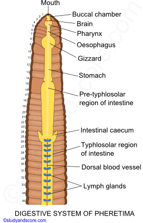
Arises from 26th segment and extends forward to 22/23
Secretes amylolytic ferment (amylase)
❖ Typhlosole:
Highly glandular, vascular, longitudinal ridge
Blind and folded structure
Increases absorption area for digested food
15 onwards
Region
Segments
Details
Pre-typhlosolar region
15–25
Typhlosolar region
27–23/25 segments in front of anus
Post-typhlosolar region (Rectum)
Last 23/25 segments
Absorption of H₂O from waste food and elimination of waste; also known as Rectum
📚
CIRCULATORY SYSTEM
▢ Blood:
Red in colour
Respiratory pigment = Haemoglobin is dissolved in the blood plasma.
So, the blood is red in colour.
No Red Blood Cells
Oxygen carrying capacity of the blood in earthworm is less than that of vertebrate blood.
▢ Blood Vessels:
1 / 2
❖ Longitudinal blood vessels:
S.N.
Dorsal Blood Vessel
Ventral Blood Vessel
Lateral Oesophageal Blood Vessels
Subneural Blood Vessel
Supra-oesophageal Blood Vessel
Location
1st to last segment
1st to last segment
1st to 13th segment on both sides of alimentary canal
14th to last segment below the ventral nerve cord
9th to 13th segment
Features
Largest blood vessel in the earthworm
Bears a pair of valves in each segment
Found below alimentary canal
Single
No valves
After 13th segment, they unite to form a single sub-neural vessel
Single
Smallest lateral blood vessel
Flow of Blood
Posterior to Anterior direction
Anterior to Posterior direction
Type
Distributing type = 1st to 13th segment
Collecting type = 14th onwards
Collects from Dorso-intestinals and Sub-neurals through Commissural vessel
Main distributing blood vessel
Gives off paired tegumentary vessels in each segment to supply blood to segmental organs
Collects blood from seminal vesicles present in 11th and 12th segments
Receives blood from latero-oesophageal vessel through anterior loops
Also receives from stomach and gizzard through ring vessel
1 segment bears about 12 ring vessels
◉ Key: One pair of Commissural vessel is present in each segment. It transfers blood from sub-neural to dorsal vessel. It gives a branch called Septo-intestinal to intestine.
❖ Transverse blood vessels:
S.N.
Lateral Hearts
Anterior Loops
Lateral Oesophageal Hearts
Location
7th and 9th segments
10th and 11th segments
12th and 13th segments
Features
One pair in each segment
Sends blood from dorsal vessel to ventral vessel
Each vessel with 4 pairs of valves
One pair in each segment
Sends blood from lateral oesophageal vessel to supra-oesophageal vessel
One pair in each segment
Sends blood from supra-oesophageal and dorsal vessel to ventral vessel
Each vessel with 3 pairs of valves
▢ Hearts:
❖ Description: Heart of earthworm is muscular, thick-walled, valvular, and pulsating.
▢ Blood Glands:
❖ Location: Segments 4, 5, and 6 above the pharyngeal mass
❖ Association: Blood glands are associated with pharyngeal nephridia
❖ Function: Manufacture of blood corpuscles and haemoglobin
▢ Lymph Glands:
❖ Location: Both sides of dorsal blood vessel from 26th segment onwards
❖ Number: 1 pair per segment
❖ Shape:
◉ Size: Small
◉ Color: Whitish
❖ Function: Supposed to produce certain phagocytic cells (Amoebocytes)
📚
RESPIRATORY SYSTEM
Cutaneous respiration
No respiratory organs
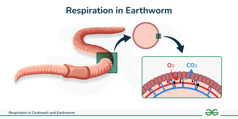
Exchange of gases takes place through moist skin
The absorptive area of earthworm is more than its volume. So, earthworm doesn't require any respiratory organ.
The cutaneous blood supply of the animal is more which is helpful in the process of gaseous exchange.
If the skin of the earthworm dries, it cannot respire. It is due to asphyxia.
100 cc of blood carries 10 cc of oxygen.
Note: Vertebrate blood carries 20 cc of oxygen per 100 cc.
📚
EXCRETORY SYSTEM
S.N.
Septal nephridia / Typical nephridia
Pharyngeal nephridia
Integumentary nephridia
1 / 2
1. Location
Attached to both faces of each intersegmental septum behind 15th segment.
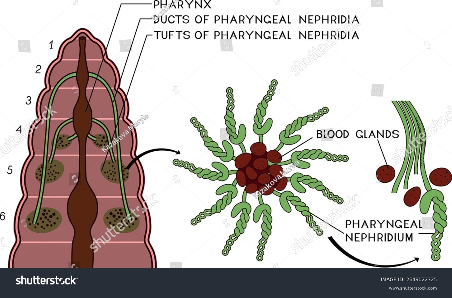
4th, 5th and 6th segments on either side of alimentary canal.
Scattered in the ventral body wall in all segments except first 2 or 3. Absent in anterior 14 segments.
2. Number
Each septum has 2 rows with 80–100 nephridia per septum.
In the form of paired tufts — 3 pairs of tufts, each containing numerous nephridia.
200–250 per segment. 10× more in clitellar region = 'Forest of nephridia'. No. of nephridiopores = No. of nephridia.
3. Type
Enteronephric nephridia
Enteronephric nephridia
Exonephric nephridia
4. Parts of Nephridia
a. Nephrostome (with upper & lower lips) b. Neck (ciliated duct) c. Main body (twisted & straight loop) d. Terminal duct (ciliated) e. Nephridial tube (single coiled tubule with 4 ciliated regions)
Without nephrostome Paired tufts with common pharyngeal duct.
Without nephrostome Smallest nephridia
5. Flow of Wastes
Nephrostome → Neck → Main body → Terminal duct → Septal excretory canal → Two supra-intestinal excretory canals → Intestine
Terminal ducts of tufts open into a common pharyngeal nephridial duct (common excretory duct)
-
6. Features
a. Largest nephridia b. Only type with nephrostome
Ducts from 4th & 5th segments open into pharynx; 6th into buccal cavity.
a. Smallest nephridia b. Most numerous c. V-shaped
📚
REPRODUCTIVE SYSTEM
▢ Male reproductive system:
Male Reproductive Parts
Number
Location
Features
Function in Development of Sperm
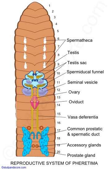
1. Testes
2 pairs
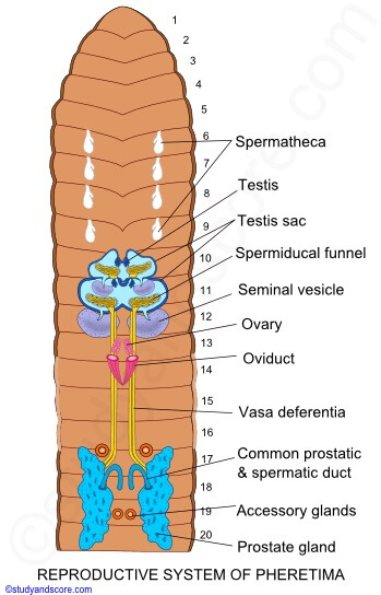
10th and 11th segments
i. Small white structure enclosed within testis sac.
ii. Segmental septa of testes are 9/10th and 10/11th segments.
iii. Each testis consists of 4–8 finger-like projections, containing spermatogonia at different stages of development.
iv. Testes remain well developed in young earthworm but degenerate in mature earthworm.
i. Produce spermatogonia.
2. Testes sacs
2 pairs
10th and 11th segments
i. Thin-walled fluid-filled membranous bag, encloses the testes.
ii. Each testis sac is ventro-lateral in position and bilobed in front.
iii. Also encloses spermiducal funnel.
iv. The testis sac of 10th and 11th segment communicate with the respective seminal vesicles of 11th and 12th segment.
i. The spermatogonia are shed into the testis sac, from where they enter into seminal vesicles for maturation.
3. Spermiducal funnels
2 pairs
10th and 11th segments
i. Funnel-like structure with folded and ciliated margin.
ii. Lies behind the testis, enclosed within testis sac.
iii. Each spermatic funnel leads to vas deferens.
i. Collects sperms.
4. Seminal vesicles
2 pairs
11th and 12th segments
i. Oval sacs, found on both sides of stomach.
ii. Developed from septa as outgrowth, so called septal pouches.
iii. 12th segment seminal vesicles are larger than 11th segment ones.
iv. Seminal fluid with fructose, the chief food for sperm.
i. Play role in maturation of spermatozoa from spermatogonia.
1. Produce sperms.
2. Sperms rest here until copulation.
5. Vasa deferentia
2 pairs
Extends from 10th/11th to 18th segment
i. Narrow elongated ducts, run on either side of alimentary canal piercing through inter-segmental septa.
ii. In 18th segment, they incorporate with prostatic duct forming common prostatic and spermatic duct, which opens outside through male genital aperture on the ventral wall.
i. Conduct sperms.
6. Prostate gland
1 pair
Extends from 16th/17th to 20th/21st segment
i. Large, elongated white coloured and irregularly-shaped lobulated gland.
ii. The prostatic secretion is useful for the activation of sperms.
i. Provides nourishment to the sperm.
7. Accessory glands and Genital Papillae
2 pairs
17th and 19th segments
i. Found on either side of the nerve cord, attached to the floor of body cavity.
ii. Secretes the sticky substance which helps in attachment of two worms during copulation.
iii. Opens out through genital papillae.
▢ Female reproductive system:
Female Reproductive Parts
Number
Location
Features
1. Ovaries
1 pair
13th segment
i. Small whitish masses on either side of ventral nerve cord.
ii. Consist of finger-like projections called ovarioles, which contain eggs of different developmental stages.
iii. The eggs at the apices are the matured ones.
2. Oviducal funnel
1 pair
13th segment just beneath ovaries
3. Oviducts
1 pair
Extending from 13th to 14th segment
i. Starts from 13th segment as oviducal funnel, meant for receiving ova from the ovary.
ii. In 14th segment, they converge and open outside ventrally through female genital pore.
4. Spermathecae
4 pairs
6th, 7th, 8th, and 9th segments ventrolaterally
i. Spermathecae (seminal receptacles) are for receiving sperm from another worm during copulation and temporary storage of sperms.
ii. Each spermatheca is flask-shaped, consisting of a pear-shaped ampulla and a short elongated diverticulum.
1. Diverticulum = Storage of sperms received from another worm during copulation.
2. Ampulla = Provides nourishment to the stored sperms.
iii. Spermathecal openings are in the intersegmental grooves of 5/6th, 6/7th, 7/8th, and 8/9th segments.