📚
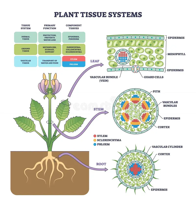
TISSUE SYSTEM
📖
Epidermal tissue system
▢ Definition: Outer covering of plants
▢ Includes:
- Epidermis
- Stomata
- Trichomes / hairs
📝
Epidermis
Root | Stem | Leaves |
|---|---|---|
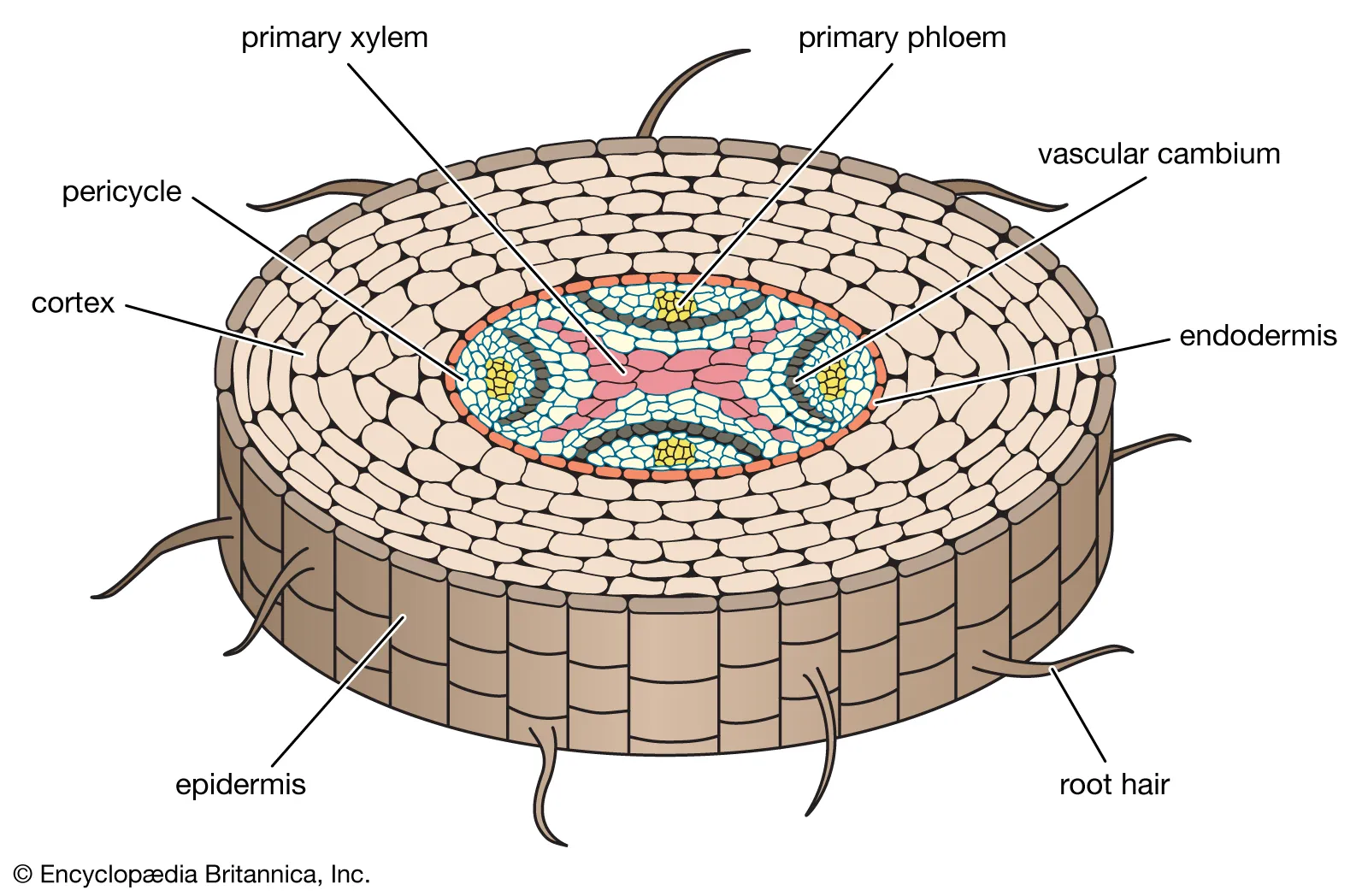 | 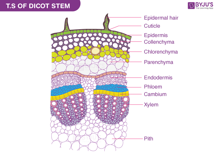 | 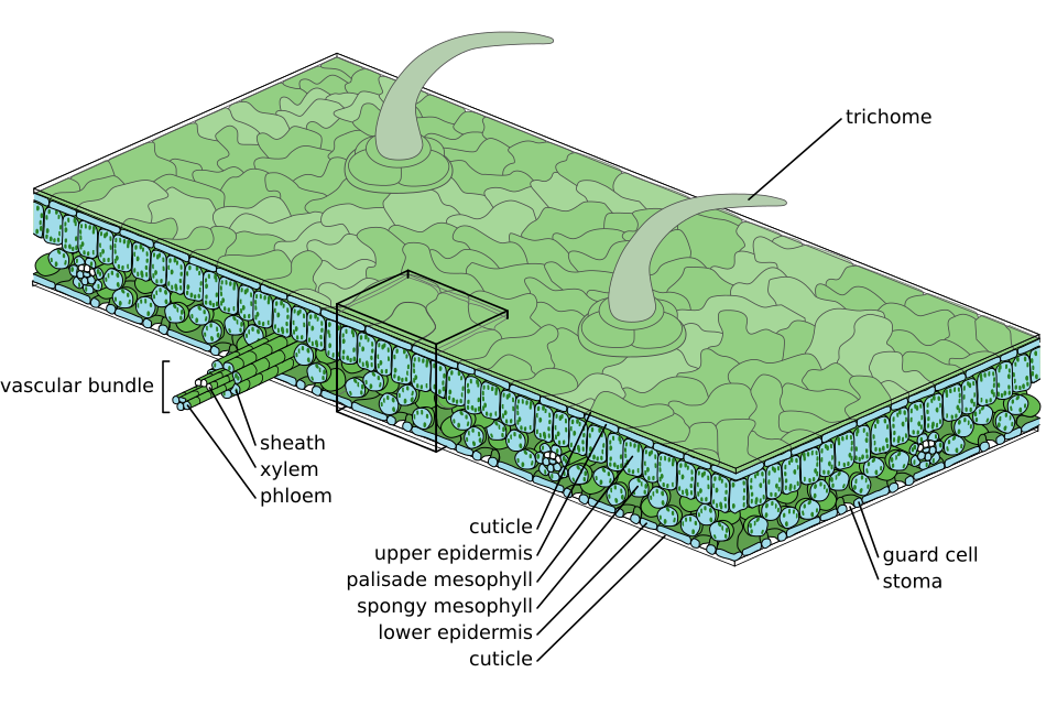 |
▢ Definition: Epi = upon; derma = skin → continuous outer layer interrupted by stomata + lenticels
▢ Layering:
Table 1: Epidermis layering
Type | Occurrence / Example |
|---|---|
Generally | Single-layered |
Multilayered epidermis | Phylloclades of Casuarina; leaves of Nerium + Ficus; hygroscopic roots of epiphytes |
Velamen | Multiple epidermis on hygroscopic roots; cells dead |
▢ Cuticle:
- •Present on epidermis of stem + leaves
- •Absent in root
- •Protection to epidermis
- •Checks excessive transpiration
▢ Chloroplast:
- •Epidermal cells living
- •Generally no chloroplast
- •Exceptions → shade plants / sciophytes, hydrophytes, some ferns
- •Guard cells contain chloroplast
- •Guard cells living but heteromorphic due to absence of RUBISCO
▢ Special Epidermal Cells / Contents:
Table 1: Special epidermal points
Structure / Content | Occurrence | Function / Note |
|---|---|---|
Bulliform / motor / bubble-like cells | Upper epidermis of isobilateral leaves / monocots | Rolling + unrolling of leaves; mainly rolling |
Epiblema / piliferous layer / rhizodermis | Outermost layer of root | Not cutinised/suberised; no stomata |
Stomata + lenticels | Epidermis | Confined to epidermis |
Silica | Epidermal cells of grasses + Equisetum | Mechanical strength |
Lithocysts | Epidermal cells containing cystoliths |
📝
Stomata
▢ Definition: Minute pores on epidermal surface of leaves + some herbaceous stems
▢ Structure:
- •Each stoma guarded by 2 guard cells
- •Guard cells = specialized epidermal cells
- •Subsidiary / accessory cells surround guard cells
▢ Guard Cell Shape:
Table 1: Guard cell shape
Plant group | Guard cell shape |
|---|---|
Dicots | Kidney-shaped / bean-shaped |
Some monocots e.g., doob grass / Cynodon dactylon, maize | Dumb-bell / barbell-shaped |
▢ Distribution:
Table 1: Stomatal distribution
Plant / Leaf type | Stomata distribution |
|---|---|
Dicot leaves | Scattered; generally lower epidermis |
Monocot leaves | Scattered in rows; both surfaces |
Free-floating hydrophytes e.g., lotus, Nymphaea | Upper epidermis |
Submerged hydrophytes | Absent / non-functional |
Algae + fungi | Totally absent |
▢ Leaves Based on Stomata:
Table 1: Leaves by stomatal distribution
Leaf type | Stomata found on | Examples |
|---|---|---|
Epistomatic | Upper surface | Free-floating hydrophytes e.g., lotus, Nymphaea |
Hypostomatic | Lower surface | Common dicot leaves |
Amphistomatic | Both surfaces | Common monocot leaves |
📝
Trichome / Hairs
Table 1: Trichomes
Type | Occurrence | Function |
|---|---|---|
Unicellular | Roots | Absorption / protection |
Multicellular | Stems | Checks excess water loss + protection |
📖
Ground tissue system
▢ Definition: Tissue system between epidermis and vascular tissue
▢ Components:
- Hypodermis
- Cortex
- Endodermis
- Pericycle
- Medulla / pith
- Medullary rays / pith rays
- Conjunctive tissue
📝
Hypodermis
Table 1: Hypodermis
Point | Data |
|---|---|
Position | Outermost layer of cortex |
Herbaceous dicot stem | Collenchymatous |
Monocot stem | Sclerenchymatous |
📝
Cortex
- •Present inside hypodermis
- •Usually parenchymatous
- •Abundant intercellular space
- •Innermost layer → endodermis
📝
Endodermis / Starch Sheath
▢ Definition: Single-layered tissue separating cortex from stele
▢ System: Part of extrastelar ground tissue system
▢ Cell Features:
- •Barrel-shaped
- •No intercellular spaces
- •Living
- •Starch-containing → starch sheath
▢ Casparian Strips:
- •Radial + tangential wall thickening; mainly radial wall
- •Made of lignin + suberin + cutin
- •Stripes/bands → Casparian strips / Casparian bands
- •Casparian strips + passage cells also found in exodermis
▢ Passage Cells:
- •Present just against protoxylem in root
- •Exodermis may occur in some roots e.g., hygroscopic roots of epiphytes
▢ Root Point: Well-developed endodermis = constant feature of roots; not constant in stem
📝
Pericycle
▢ Position: Inside endodermis
▢ Layers: Single-layered or few-layered
▢ Cell Type: Thin-walled parenchymatous or rarely thick-walled sclerenchymatous
▢ Occurrence:
- •Present in most roots
- •Absent in roots of parasitic plants + hydrophytes
▢ Special Cases:
Table 1: Pericycle special cases
Plant / Case | Pericycle feature |
|---|---|
Smilax root / monocot | Multilayered + sclerenchymatous |
Sunflower stem | Heterogeneous → thick-walled + thin-walled cells |
▢ Functions:
Table 1: Pericycle functions
Pericycle type | Function |
|---|---|
Thick-walled | Mechanical |
Thin-walled | Storage |
Dicot root pericycle | Can form cambium |
📝
Medulla / Pith and Medullary Rays
▢ Definitions:
Table 1: Pith and pith rays
Term | Meaning |
|---|---|
Medulla / pith | Core of plant axis occupied by parenchymatous tissue |
Medullary rays / pith rays | Pith radiating between vascular bundles |
▢ Functions:
Table 1: Functions
Structure | Function |
|---|---|
Pith | Food storage |
Medullary rays | Lateral transportation |
▢ Distribution:
Table 1: Pith distribution
Plant part | Pith status |
|---|---|
Dicot stem | Distinct / developed |
Monocot root | Distinct / developed |
Monocot stem | Scattered |
Dicot root | Indistinct / small / absent |
Triticum stem | Central region hollow → pith cavity |
Ricinus | Pith cavity among dicots |
📝
Conjunctive Tissue
Parenchymatous tissue between xylem and phloem bundles
📝
Cortex and Stele
Table 1: Cortex vs stele
Region | Meaning |
|---|---|
Cortex | Ground tissue outside endodermis |
Stele | All tissues inside endodermis |
Monocot stele | Atactostele |
Dicot stele | Eustele |
Endodermis absent in monocot stem → ground tissue not differentiated into cortex and stele
📝
MCQ Point
📖
Vascular tissue system
▢ Definition:
- •Central column of root/stem axis = stele
- •Stele made of vascular bundles
- •Stelar region includes all tissues inside pericycle
- •Vascular bundle = xylem + phloem + cambium if present
- •Cambium = strip of primary meristem
▢ Types of Vascular Bundles:
❖ Radial:
- •Xylem + phloem in separate bundles
- •Present on different radii
- •Alternate arrangement
- •Equal in number
- •Example → roots of dicots + monocots
❖ Conjoint:
▢ Definition: Xylem + phloem in same bundle and same radius
▢ Example: Stem
▢ Types:
❖ Collateral:
Table 1: Collateral vascular bundle
Type | Cambium | Example / Note |
|---|---|---|
Open collateral | Present between xylem + phloem | Dicot stem; conjoint, collateral, open |
Closed collateral | Absent | Monocot stem; conjoint, collateral, closed |
Phloem outside/pericycle side; xylem towards centre/pith
❖ Bicollateral:
▢ Definition: Phloem present on both sides of xylem
▢ Bundle Nature: Always open
▢ Arrangement: Phloem → cambium → xylem → cambium → phloem
▢ Examples:
- •Stem of Cucurbitaceae
- •Stem of Solanaceae
- •Stem of Convolvulaceae
▢ MCQ Point:
❖ Concentric:
▢ Definition: Xylem + phloem arranged in definite circle
▢ Nature: Always closed
▢ Occurrence: Characteristic of pteridophytes + few monocots
▢ Types:
Table 1: Concentric vascular bundles
Type | Arrangement | Examples |
|---|---|---|
Amphicribal / hadrocentric | Xylem central; phloem surrounds xylem | Ferns, some aquatic angiosperms, Lycopodium, Selaginella |
Amphivasal / leptocentric | Phloem central; xylem surrounds phloem | Underground stem of some monocots e.g., dagger plant / Yucca, dragon plant / Dracaena, sweet flag / Acorus |
📚
ROOT-STEM TRANSITION
- •Conversion: radial + exarch vascular bundle → conjoint + endarch vascular bundle
- •Xylem swings during transition
- •Phloem remains stationary
- •Transition zone present in hypocotyl
📚
ORIGIN OF LATERAL ROOTS AND LATERAL BRANCHES OF STEM
Table 1: Origin of lateral organs
Structure | Origin | Source / Seat |
|---|---|---|
Lateral root | Endogenous | Pericycle |
Seat of lateral root origin | Just against protoxylem | Pericycle opposite protoxylem |
Lateral branch of stem | Exogenous | Outer part of cortex |
📚
ANATOMY OF STEM
▢ Dicot Stem vs Monocot Stem:
Table 1: Anatomy of dicot and monocot stem
Feature | Dicot stem | Monocot stem |
|---|---|---|
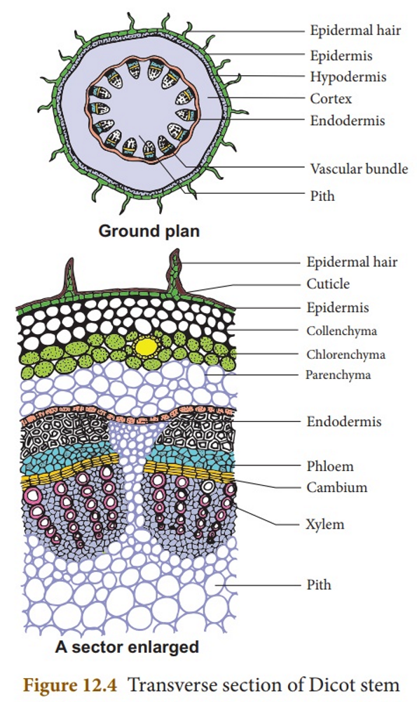 | 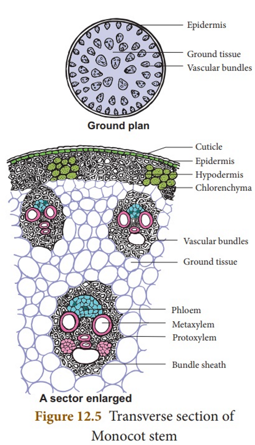 | |
Epidermis | Epidermis with multicellular hairs + stomata with kidney-shaped guard cells | Outer epidermis cutinized; stomata with dumb-bell-shaped guard cells |
Hypodermis | Collenchymatous | Sclerenchymatous |
Internal tissue arrangement | Concentric | Concentric arrangement absent |
Ground tissue | Differentiated into hypodermis, general cortex, endodermis, pericycle, pith | Not differentiated |
Stem centre | Almost always solid | Usually hollow centre; exception maize |
Vascular bundle arrangement | Ring around pith | Scattered throughout ground tissue |
Vascular bundle number/size | Few + similar size | Numerous; smaller at periphery, larger towards central axis |
Vascular bundle type | Conjoint, collateral, open | Conjoint, collateral, closed |
Bundle sheath | Absent | Sclerenchymatous bundle sheath present |
Bundle outline | Wedge-shaped | Oval / rounded |
Phloem parenchyma | Present | Absent |
Protoxylem cavity | Absent | Present; exception Asparagus |
Vessel arrangement | Rows/chains; linear fashion | Y-shaped manner |
Stele | Eustele / dictyostele | Atactostele; most advanced stele |
Pericycle | Alternating zones of parenchyma + sclerenchyma | Mostly absent |
Medullary rays | Present | Absent |
Secondary growth | Present | Absent |
📚
ANATOMY OF ROOT
▢ Common Features of Dicot and Monocot Roots:
- •Wide cortex made of thin-walled cells
- •Vascular bundle radial
- •Xylem exarch
- •Distinct endodermis + passage cells
- •Lateral roots arise opposite protoxylem points from pericycle → endogenous
▢ Dicot Root vs Monocot Root:
Table 1: Anatomy of dicot and monocot root
Feature | Dicot root | Monocot root |
|---|---|---|
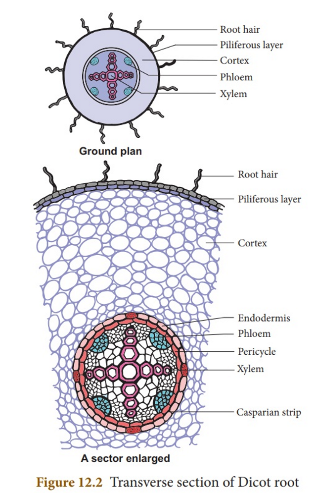 | 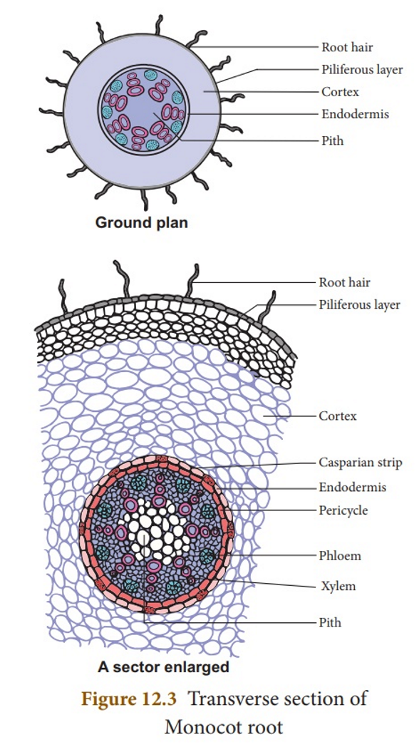 | |
Cortex | Narrow | Very wide |
Outer tissues during secondary growth | Epiblema, cortex, endodermis peeled off and replaced by cork | Cork not formed; only epiblema peeled off; cortex + endodermis persist |
Endodermis | Less thickened; Casparian strips prominent | Casparian strips visible only in young roots; later endodermal cells highly thickened |
Passage cells | Generally absent | Generally present in endodermis opposite protoxylem |
Pericycle produces | Lateral roots + cork cambium + vascular cambium | Lateral roots only |
Xylem and phloem bundles | ||
Conjunctive tissue | Parenchymatous | Parenchymatous or sclerenchymatous |
Conjunctive parenchyma | Forms cambium | Does not form cambium |
Pith | Small or absent | Well-developed |
Protoxylem and metaxylem | Linear fashion | Linear or V-shaped |
Secondary growth | Present | Absent |
▢ Special Points:
- •Grafting not possible in monocots → cambium absent
- •Girdling not possible due to scattered vascular bundles
📚
HISTOLOGY OF LEAF
▢ General Points:
- •Dicot leaf horizontal; upper sun-facing part darker → dorsiventral leaf
- •Monocot leaf nearly vertical; both surfaces equally exposed → isobilateral leaf
- •Leaf anatomy → upper epidermis + lower epidermis + mesophyll + vascular bundle
- •Inverted vascular bundle → xylem upper side, phloem lower side
▢ Vascular Bundle:
- •Conjoint
- •Collateral
- •Mesarch xylem
▢ Dicot Leaf vs Monocot Leaf:
Table 1: Histology of dicot and monocot leaf
Feature | Dicot leaf | Monocot leaf |
|---|---|---|
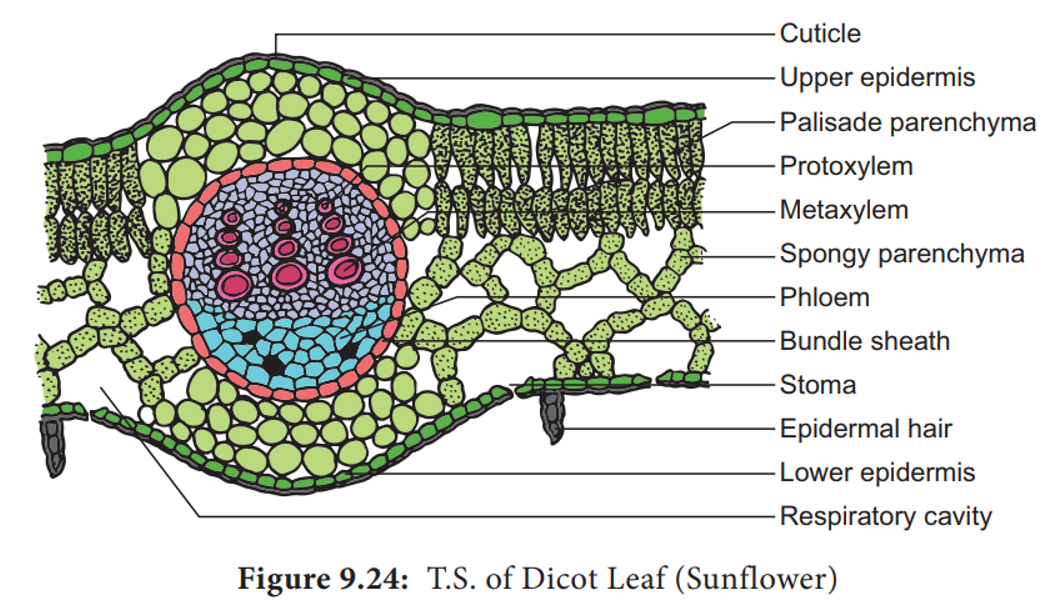 | 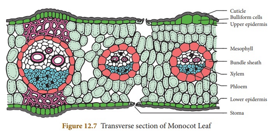 | |
Cuticle thickness | Upper epidermis thicker than lower epidermis | Same in both epidermis |
Bulliform / motor cells | Absent | Present |
Number of stomata | Greater on lower surface than upper | Equal on both surfaces |
Guard cells | Kidney-shaped | Dumb-bell-shaped |
Mesophyll | Differentiated into upper tightly packed palisade parenchyma + lower loosely arranged spongy parenchyma | Not differentiated into palisade and spongy parenchyma |
Bundle sheath extension | Present in larger veins; parenchymatous | Present; sclerenchymatous |
Bundle sheath cells | Colourless | |
Lysigenous cavity in xylem | Absent | Usually present |
Q1.
Plasmolysis in plant cells occur when they are placed in: [IOM 1998]
📅IOM 1998
Q2.
Fertilizers can kill plants due to: [IOM 1996]
📅IOM 1996
Q3.
When the fluid outside a cell has greater concentration of a given molecule than the fluid inside the cell, the external fluid is: [BPKIHS 2004]
📅BPKIHS 2004
Q4.
The process of imbibition can be noticed when: [BPKIHS 2004]
📅BPKIHS 2004
Q5.
An Amoeba survives in a hypotonic solution that destroys a human RBC. It is due to [BPKIHS 2004]
📅BPKIHS 2004
Q6.
Imbibition demonstrates the process of: [BPKIHS 2006]
📅BPKIHS 2006
Q7.
When a cell is fully turgid its: [BPKIHS 1997]
📅BPKIHS 1997
Q8.
The turgidity of the cell is maintained by: [MOE 2008]
📅MOE 2008
Q9.
Plasmolysis is defined as: [MOE 2063]
📅MOE 2063
Q10.
Selective permeability identifies the process of: [MOE 2062]
📅MOE 2062
Q11.
Imbibition theory for ascent of sap was given by: [MOE 2000]
📅MOE 2000
Q12.
Plant absorb water by: [MOE 2065]
📅MOE 2065
Q13.
Transpiration is high during following condition: [IOM 2007]
📅IOM 2007
Q14.
Potometer is used to measure: [IOM 2006]
📅IOM 2006
Q15.
Out of water absorbed by plants, water actually used by them is: [IOM 1997]
📅IOM 1997
Q16.
Opening of the stomata is because of: [IOM 1997]
📅IOM 1997
Q17.
K+ exchange theory is concerned with: [IOM 1997]
📅IOM 1997
Q18.
Transpiration is highest for ……… transpiration: [IOM 1996]
📅IOM 1996
Q19.
Loss of water in the form of vapour from the living tissue of aerial parts of the plant is termed as: [BPKIHS 2006]
📅BPKIHS 2006
Q20.
Rate of transpiration is affected by humidity in: [BPKIHS 2000]
📅BPKIHS 2000
Q21.
The pores specialized for guttation in plants are called: [BPKIHS 2001]
📅BPKIHS 2001
Q22.
Cells in the root having power of division: [BPKIHS 2003]
📅BPKIHS 2003
Q23.
Which one keeps its stomata open during night and closed during day? [BPKIHS 2004]
📅BPKIHS 2004
Q24.
The rate of transpiration during storm is [BPKIHS 1995]
📅BPKIHS 1995
Q25.
There is accumulation of water drops at the leaf margin of Colocasia early in the morning, this is the result of [BPKIHS 1996]
📅BPKIHS 1996
Q26.
If a plant kept in air free of CO_2 [BPKIHS 1998]
📅BPKIHS 1998
Q27.
The translocation of food in plants occurs in the form of: [IE 2001]
📅IE 2001
Q28.
Who proposed the 'Cohesion-Tension' and 'Transpiration pull theory'? [MOE 2062]
📅MOE 2062
Q29.
Root pressure is maximum when: [MOE 2062]
📅MOE 2062
Q30.
The nature of dry cobalt chloride paper which is used in transpiration is: [MOE 2058]
📅MOE 2058
Q31.
Minute pore-like opening found in cork that help in exchange of gases are called: [MOE 2052]
📅MOE 2052
Q32.
The region just behind the root apex is: [MOE 2061]
📅MOE 2061
Q33.
The maximum amount of solute present in the phloem sap is: [IE 2002]
📅IE 2002
Q34.
In roots, the tissue which helps in the absorption of water and minerals is: [IE 2002]
📅IE 2002
Q35.
Root tip is more distant than any other apical part of plant due to: [IE 2005]
📅IE 2005
Q36.
The lowest water potential in the xylem are in the: [IE 2007]
📅IE 2007
Q37.
Cell wall enlarges if kept in: [IE 2008]
📅IE 2008
Q38.
Chlorophyll contains which metal ion: [IE 2008]
📅IE 2008
Q39.
A micronutrient absorbed by foliage is [IE 2009]
📅IE 2009
Q40.
The immediate effect of destruction of ATP inside cell is in [BPKIHS 2016]
📅BPKIHS 2016
Q41.
When a plant cell is kept in water, water continues to move inside until the [BPKIHS 2016]
📅BPKIHS 2016
Q42.
Bacteria can't grow in honey because [BPKIHS 2016]
📅BPKIHS 2016
Q43.
Common disease caused due to deficiency of P, K, Mg, Mn, Ca "and" N is [BPKIHS 2014]
📅BPKIHS 2014
Q44.
Which of the following becomes difficult in absence of ATP? [BPKIHS 2012]
📅BPKIHS 2012
Q45.
Which of the following factors does not affect osmosis? [BPKIHS 2010]
📅BPKIHS 2010
Q46.
The device which is used to nullify the effect of gravity is called [IOM 2014]
📅IOM 2014
Q47.
Plasmolysis is caused when cell is placed in [IOM 2016]
📅IOM 2016
Q48.
Site of evaporation of water is [IOM 2012]
📅IOM 2012
Q49.
For plasmolysis, cell is placed in [IOM 2012]
📅IOM 2012
Q50.
The cell becomes turgid when placed in [MOE 2014]
📅MOE 2014
Q51.
Ascent of sap in plants is due to [MOE 2014]
📅MOE 2014
Q52.
Guttation is the manifestation of [MOE 2012]
📅MOE 2012